


🩺 AI in Medical Imaging for Cancer: How New Models Are Changing Radiology, Pathology, and Patient Outcomes

Sharing is SO MUCH APPRECIATED!

Last updated: June 10, 2026
Quick Answer
AI in medical imaging for cancer is already active in clinical settings worldwide, helping radiologists and pathologists detect tumors earlier, reduce missed diagnoses, and process scans faster than traditional workflows allow. A meta-analysis of 34 studies found that AI assistance improved radiologist sensitivity from 0.67 to 0.79 and specificity from 0.82 to 0.87 [3]. The technology is not replacing doctors — it is giving them a sharper set of tools.
Key Takeaways
- AI-assisted cancer imaging has shown measurable improvements in detection rates for colorectal, esophageal, and prostate cancers across multiple randomized controlled trials [1]
- AI improves both sensitivity (catching more true positives) and specificity (reducing false alarms) when used alongside human radiologists [3]
- Deep learning models can analyze MRI, CT, PET, and whole-slide pathology images to flag lesions, segment tumors, and extract radiomic features automatically [4]
- GPU-accelerated AI platforms have cut operational costs by up to 85% in some cancer genomics applications [5]
- AI is especially promising for rural and underserved communities where specialist radiologist access is limited
- Current AI tools are strongest in breast, lung, colorectal, and prostate cancer screening; evidence is weaker for gastric and liver cancers [1]
- Insurance coverage for AI-assisted imaging varies widely and is still evolving in Canada, the US, and elsewhere
- Over-reliance on AI without physician oversight remains a real clinical risk
- Standardization of imaging data and scanner calibration are active challenges limiting wider AI deployment [9]
- The pace of improvement in AI cancer imaging is accelerating, with multimodal models now integrating radiology, pathology, and genomics data together [2]

What Exactly Is AI Doing in Medical Imaging for Cancer Detection
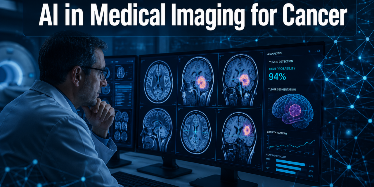
AI in cancer imaging works by training algorithms — most often deep learning neural networks — on large datasets of labeled medical images. The models learn to recognize patterns associated with malignancy: irregular borders, unusual density, abnormal cell structures. Once trained, they can analyze new scans and flag areas of concern for a clinician to review.
In practice, AI is being used for several distinct tasks:
- Detection: Identifying suspicious lesions or nodules on CT, MRI, or X-ray scans
- Segmentation: Outlining the exact boundaries of a tumor to assist in treatment planning
- Classification: Distinguishing benign from malignant findings
- Risk stratification: Estimating how likely a finding is to be cancer based on imaging features
- Radiomic feature extraction: Pulling quantitative data from images that the human eye cannot reliably measure [4]
In pathology, AI analyzes digitized tissue slides (whole-slide images) to identify cancerous cells, grade tumors, and detect subtle patterns linked to treatment response [7].
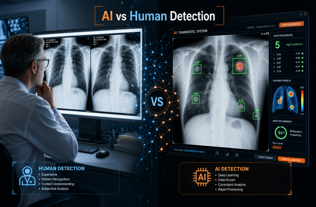
How Accurate Are AI Models Compared to Human Radiologists
AI models do not consistently outperform experienced radiologists working alone, but they reliably improve performance when the two work together. A systematic review of 34 studies showed that AI assistance raised sensitivity from 0.67 to 0.79 and specificity from 0.82 to 0.87 — a meaningful gain in both catching cancer and avoiding unnecessary follow-up [3].
The key insight: AI works best as a second reader, not a replacement. It catches cases a tired or overloaded radiologist might miss, and it flags low-confidence reads for extra scrutiny.
Important caveat: Half of the studies in that same review had concerns about bias in their design [3]. Real-world performance can differ from controlled study conditions.
What Types of Cancer Are AI Imaging Models Best at Detecting
AI cancer imaging tools show the strongest evidence in colorectal, esophageal, prostate, breast, and lung cancers. A review of 49 randomized controlled trials found AI improved adenoma detection in colorectal cancer (pooled relative risk of 1.22) and showed similar gains in esophageal and prostate cancer screening [1]. For gastric and liver cancers, the evidence so far is less compelling [1].
Choose AI-assisted screening if: The cancer type has large, well-labeled imaging datasets available and a defined screening pathway (breast mammography, lung CT, colonoscopy-based colorectal screening).
Be more cautious if: The cancer type has limited training data or highly variable imaging presentation.
How Does AI Help in Early Stage Cancer Detection
Early detection is where AI has the greatest potential impact on patient outcomes. Small tumors and pre-cancerous changes are easy to miss on routine scans, especially when a radiologist is reading hundreds of images per shift. AI models trained on thousands of confirmed early-stage cases can flag subtle findings that fall below the threshold of human attention.
In tissue imaging, AI has been developed for regression, classification, segmentation, and generation tasks on histopathological images, improving the accuracy of early cancer identification before symptoms appear [7]. For lung cancer, AI tools applied to low-dose CT scans can identify sub-centimeter nodules and estimate malignancy probability, giving clinicians a head start on follow-up decisions.
Thinking about healthy aging and preventive health? Earlier cancer detection through AI-assisted screening is one of the most direct ways technology is extending quality of life.
What Kind of Medical Imaging Data Do These AI Models Use
AI cancer models are trained on multiple imaging modalities, including:
- CT (computed tomography): Lung, colon, kidney cancers
- MRI (magnetic resonance imaging): Brain, breast, prostate cancers
- PET scans: Metabolic activity mapping for staging
- Mammography: Breast cancer screening
- Whole-slide pathology images: Tissue biopsy analysis for nearly all cancer types
- Chest X-rays: Lung abnormality triage
Next-generation models go further, integrating imaging data with genomics and electronic health records to build a fuller picture of each patient’s cancer biology [2]. An autonomous AI system deployed across 17 major healthcare systems in India processed over 150,000 chest X-rays, achieving precision up to 98% and recall over 95% for multiple pathologies [8].
Standardization matters here. Variations between scanners can distort AI outputs. Physical color calibration of digital pathology scanners has been shown to improve AI model performance in Gleason grading for prostate cancer, making results more reliable across different clinical settings [9].
Can AI Miss Cancer Signs That a Human Doctor Might Catch
Yes. AI models have known blind spots, and this is a real clinical concern. Models trained on one population or scanner type can perform poorly when applied to a different demographic or equipment setup. They can also miss rare cancer presentations that were underrepresented in training data.
Common failure modes include:
- Distribution shift: The real-world scan looks different from training data (different scanner, patient population, or image quality)
- Rare subtypes: Unusual cancer morphologies that appear infrequently in training sets
- Contextual gaps: AI does not read a patient’s clinical history, symptoms, or lab results the way a physician does
This is why every major clinical AI deployment pairs the model with a qualified physician who makes the final call. AI flags; doctors decide.
What Are the Risks of Relying Too Much on AI in Cancer Diagnosis
Over-reliance on AI in cancer diagnosis carries several documented risks. Automation bias — the tendency to accept AI output without critical review — can cause clinicians to miss errors the model makes. If an AI system flags a scan as clear, a distracted reader may not look closely enough.
Other risks include:
- False negatives: AI misses a cancer, and the clinician trusts the result
- False positives: AI over-flags benign findings, leading to unnecessary biopsies or patient anxiety
- Equity gaps: Models trained on non-diverse datasets may perform worse for certain ethnic groups or body types
- Accountability gaps: When AI and human judgment conflict, it is not always clear who bears responsibility for the outcome
The broader societal conversation about AI accountability in healthcare is still developing, and regulatory frameworks in Canada and internationally are catching up.
How Much Does an AI Medical Imaging Scan Cost
There is no single price for an AI-assisted cancer imaging scan because the AI component is typically embedded in the radiologist’s workflow rather than billed separately. In most Canadian and publicly funded health systems, patients do not pay extra for AI tools used during their scan interpretation — the cost is absorbed by the hospital or imaging centre.
In private or US-based settings, AI-assisted reads may add a modest fee, but this varies widely by provider and scan type. GPU-accelerated AI platforms have reduced operational costs by up to 85% in some cancer diagnostics applications [5], which suggests the technology can lower costs over time rather than raise them for patients.
Bottom line: Ask your imaging provider directly whether AI tools are used in reading your scans, and whether there is any additional charge.
Is AI Medical Imaging Covered by Health Insurance
Coverage is inconsistent and evolving. In Canada, provincial health plans generally cover the imaging procedure itself (CT, MRI, mammography) without distinguishing whether AI tools were used in interpretation. In the United States, some AI-assisted mammography reads have received CPT billing codes, but coverage by private insurers varies.
As of 2026, no major health insurer has a blanket policy covering or excluding AI-assisted cancer imaging as a standalone benefit. Patients in publicly funded systems are unlikely to face out-of-pocket costs tied to AI use. Those in private insurance markets should check with their provider.
Can AI Medical Imaging Help Patients in Rural or Underserved Areas
This is one of the most compelling applications of AI in cancer imaging. Rural and remote communities — including many in northern Ontario and across Southern Georgian Bay communities — often lack on-site specialist radiologists. AI tools can pre-screen scans, prioritize urgent cases, and support general practitioners who must interpret imaging without specialist backup.
The autonomous chest X-ray AI deployed across 17 Indian healthcare systems is a direct example: it processed 2,000 scans daily across facilities that would otherwise face significant delays [8]. Similar models are being piloted in underserved Canadian regions to reduce wait times for cancer reads.
For communities where staying safe and accessing timely healthcare are ongoing concerns, AI-assisted triage could be a meaningful equalizer.
What Are the Limitations of Current AI Cancer Imaging Technologies
Despite real progress, current AI cancer imaging tools have meaningful limitations:
- Narrow training data: Most models are trained on datasets from large academic medical centres, which may not reflect community hospital populations
- Single-modality focus: Many tools analyze one scan type; integrating CT, MRI, and pathology together is still emerging [2]
- Regulatory lag: Health Canada and the FDA are still developing frameworks for AI medical device approval and post-market monitoring
- Explainability: Many deep learning models cannot clearly explain why they flagged a finding, which limits clinician trust
- Bias concerns: 50% of studies in a key systematic review had bias-related concerns [3], suggesting published accuracy figures may be optimistic
How Quickly Are AI Medical Imaging Technologies Improving
The pace is fast. GPU-accelerated computing has delivered up to 65x speed improvements in cancer genomics applications [5], and multimodal AI models that combine radiology, pathology, and genomic data are moving from research labs into early clinical trials [2]. Standardization efforts — like physical color calibration for pathology scanners — are addressing the consistency problems that held earlier models back [9].
The field is also moving from single-disease tools to broad triage platforms capable of detecting multiple pathologies in one pass [8]. Expect significant capability gains over the next two to three years as training datasets grow and regulatory pathways become clearer.
For readers interested in the intersection of technology and society, AI in cancer imaging is one of the clearest examples of how machine learning is moving from theory into everyday clinical practice.
Conclusion
AI in medical imaging for cancer is not a future promise — it is a present reality changing how radiologists and pathologists work every day. The evidence shows real gains in detection accuracy, faster triage, and new possibilities for underserved communities. But the technology works best as a tool in skilled hands, not a replacement for clinical judgment.
Actionable next steps for patients and families:
- Ask your doctor or imaging centre whether AI-assisted reading is part of your cancer screening pathway
- If you are in a rural area, ask whether AI triage tools are available to speed up specialist review of your scans
- Stay informed: AI cancer imaging is improving rapidly, and new screening options may become available through your provincial health plan
- Advocate for diverse, representative training datasets in AI tools — better data means better outcomes for everyone
For anyone navigating cancer screening or diagnosis, understanding how these tools work puts you in a stronger position to ask the right questions and make informed decisions alongside your care team.
FAQ
What is AI doing in cancer screening right now?
AI is analyzing CT, MRI, mammography, and pathology slides to flag suspicious findings, segment tumors, and prioritize urgent cases for radiologist review. It is active in clinical settings at major hospitals worldwide as of 2026.
Does AI replace radiologists in cancer diagnosis?
No. AI acts as a second reader or triage tool. A licensed physician makes the final diagnostic decision in all clinical deployments.
Which cancers benefit most from AI imaging?
Colorectal, breast, lung, esophageal, and prostate cancers have the strongest evidence base for AI-assisted imaging. Gastric and liver cancer tools are less proven [1].
Is AI cancer imaging available in Canada?
Yes, at major cancer centres and some community hospitals. Availability varies by province and facility. Rural access is growing but remains uneven.
Can AI catch cancers earlier than a human radiologist?
In some cases, yes. AI can detect sub-millimeter nodules and subtle tissue changes that fall below the threshold of reliable human detection, particularly in lung and colorectal screening.
What happens if AI and the radiologist disagree?
The radiologist’s judgment takes precedence. Many systems flag disagreements for a second human review rather than defaulting to the AI output.
How is patient data protected when AI analyzes scans?
AI cancer imaging systems in regulated healthcare settings must comply with PIPEDA (Canada) or HIPAA (US) privacy rules. Scans are typically de-identified before use in AI training.
Are AI imaging tools biased against certain patient groups?
Bias is a documented concern. Models trained on non-diverse datasets can perform less accurately for certain ethnic groups or body types. Researchers and regulators are actively working to address this.
How fast can AI read a cancer scan?
Processing time varies by system and scan complexity, but AI can analyze a chest X-ray in seconds. One deployed system averaged 2,000 chest X-rays per day across multiple hospitals [8].
Will AI lower the cost of cancer screening?
Potentially yes. GPU-accelerated AI platforms have reduced operational costs by up to 85% in some applications [5], and faster triage reduces downstream costs from delayed diagnosis.
References
[1] S1546144025006507 – https://www.sciencedirect.com/science/article/pii/S1546144025006507
[2] Artificial Intelligence Driven Cancer Diagnostics – https://aacrjournals.org/cancerres/article/85/13/2356/763104/Artificial-Intelligence-Driven-Cancer-Diagnostics
[3] PubMed – AI in Radiology Meta-Analysis – https://pubmed.ncbi.nlm.nih.gov/42064000/
[4] PMC12070983 – https://pmc.ncbi.nlm.nih.gov/articles/PMC12070983/
[5] PubMed – GPU-Accelerated AI in Cancer Diagnosis – https://pubmed.ncbi.nlm.nih.gov/40849689/
[6] arXiv – AI in Tumor Subregion Analysis – https://arxiv.org/abs/2103.13588
[7] arXiv – AI in Tissue Imaging for Early Cancer Detection – https://arxiv.org/abs/2306.16989
[8] arXiv – Autonomous AI for Multi-Pathology Detection – https://arxiv.org/abs/2504.00022
[9] arXiv – Color Calibration in Digital Pathology – https://arxiv.org/abs/2307.05519
Sharing is SO MUCH APPRECIATED!


